TMS Buran

Neural Health Blog

Latest News & Research Medical Insights
Neuronavigation for rTMS

Why Neuronavigation Matters for Repetitive TMS in Clinical Practice

Repetitive transcranial magnetic stimulation (rTMS) is an established clinical intervention, but its effectiveness depends on precision and reproducibility.

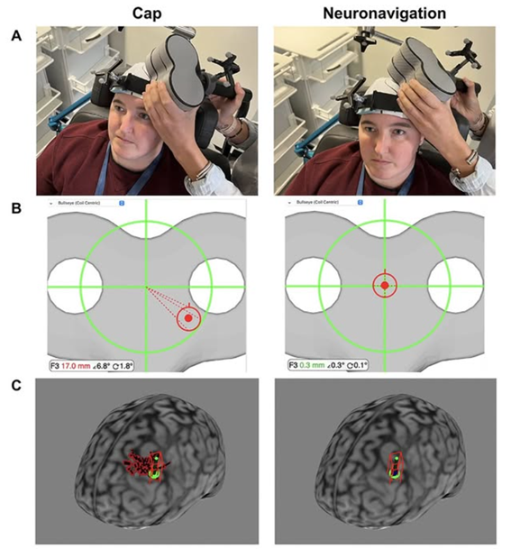
Neuronavigation represents a significant advance over traditional scalp-based targeting methods. By using each patient’s structural MRI, neuronavigation enables anatomically accurate targeting. This approach accounts for interindividual variability in skull anatomy, cortical folding, and brain–scalp distance. As a result, stimulation is delivered to the intended cortical region rather than an approximate scalp location.

Neuronavigation also improves session-to-session reproducibility. In repetitive protocols, consistent coil placement across days or weeks is critical for therapeutic efficacy. Millimetric accuracy ensures reliable engagement of the same neural substrate over time. Another key advantage is the reduction of operator-dependent variability. Landmark- or cap-based methods are sensitive to clinician experience and positioning errors. Neuronavigation standardizes targeting across operators and clinical settings. In addition, precise control of coil orientation relative to cortical gyri optimizes the induced electric field. This increases physiological specificity and may enhance clinical outcomes.

Finally, neuronavigation improves safety, documentation, and quality control. Real-time feedback and detailed logs support traceability and facilitate the integration of clinical care with research-driven protocols.

TMS Buran team

PTNS Stimulation Device
PTNS Electrode Placement

Posterior Tibial Nerve Stimulation in Chronic Pelvic Pain: Where Are We Now?

Chronic pelvic pain (CPP) is a complex, multifactorial condition affecting both men and women and significantly impairing quality of life. Its prevalence in adult women alone is estimated to range between 6% and 27%, often accompanied by urinary, gastrointestinal, sexual, and musculoskeletal symptoms, as well as anxiety and depression.

Posterior tibial nerve stimulation (PTNS/TTNS) has emerged as a low-cost, minimally invasive, and safe neuromodulation technique within this challenging clinical landscape. The posterior tibial nerve, originating from L4–S3, provides a unique anatomical gateway to modulate vesical and pudendal pathways through percutaneous or transcutaneous stimulation at the retromalleolar level.

From a neurophysiological perspective, tibial nerve stimulation is supported by the gate control theory of pain, activating large-diameter afferent fibers that inhibit nociceptive transmission at both spinal and supraspinal levels. This mechanism is particularly relevant in CPP, where central sensitization is frequently present.

Recent evidence suggests a signal of clinical benefit, especially when stimulation is integrated into a multimodal treatment approach. In male chronic prostatitis/chronic pelvic pain syndrome (CP/CPPS), comparative data indicate that percutaneous PTNS provides greater improvements in pain and quality of life than transcutaneous TTNS. In interstitial cystitis/bladder pain syndrome (IC/BPS), pilot and observational studies report improvements in pain and urinary symptoms, although methodological limitations remain.

Notably, the strongest current evidence for analgesia comes from randomized, sham-controlled trials of TTNS in primary dysmenorrhea, showing significant reductions in pain intensity and NSAID consumption after approximately 12 weekly sessions of 30 minutes.

Overall, tibial nerve stimulation stands out for its excellent safety profile, ease of implementation, and compatibility with other therapies. While it is not yet supported by large, definitive trials focused on pain as a primary outcome, it represents a promising neuromodulatory option for selected patients with chronic pelvic pain.

Dr. Öznur Buran

Dr. Öznur Buran with TMS equipment at TMS Buran Clinic

Transcranial Magnetic Stimulation and Stroke Rehabilitation

Stroke remains a leading cause of long-term disability worldwide, frequently resulting in motor dysfunction, aphasia, and chronic pain. In recent years, non-invasive brain stimulation (NIBS)—and particularly transcranial magnetic stimulation (TMS)—has gained attention as an adjuvant tool in neurorehabilitation.

TMS is based on electromagnetic induction, generating brief magnetic fields of up to 1–2.5 Tesla that induce electric currents in cortical tissue. These currents modulate neuronal excitability and network dynamics in a focal and reversible manner. Since its first description in the mid-1980s, TMS has evolved from a diagnostic method to a therapeutic neuromodulation technique.

In stroke rehabilitation, repetitive TMS (rTMS) can be delivered at high frequency (HF-rTMS) to increase cortical excitability or low frequency (LF-rTMS) to inhibit maladaptive hyperexcitability. Clinical trials have shown that both strategies can improve motor recovery. For example, increases in Fugl–Meyer motor scores have been reported in early post-stroke patients, with effects persisting at 3-month follow-up, especially when rTMS is combined with physical therapy.

Approximately one-third of stroke survivors develop aphasia, profoundly affecting quality of life. TMS and tDCS have emerged as promising adjuncts to speech therapy, targeting perilesional and contralesional language networks to facilitate functional reorganization.

Preliminary data from an open pilot study led by Dr. Öznur Buran at the Hospital Universitario de Donostia (Spain) including 40 patients—of whom 10 presented post-stroke pain, 6 aphasia with pain, and 4 upper-limb paresis with pain—demonstrate that 10 TMS sessions were well tolerated, with a low rate of adverse effects. Importantly, pain intensity decreased from an average of 1.67 pre-treatment to 0.83 post-treatment, alongside improvements in motor and language measures.

Dr. Öznur Buran

Dr. Buran and Professor Walter Frontera

Dr. Buran met Professor Walter Frontera at the 19th World Congress of the International Society of Physical and Rehabilitation Medicine (Marrakech)

About Professor Walter R. Frontera, MD, PhD:

Walter R. Frontera, MD, PhD, is Professor in the Department of Physical Medicine, Rehabilitation, and Sports Medicine at the University of Puerto Rico (UPR). He completed his medical studies and a residency in PM&R at the UPR and a doctoral degree in exercise physiology at Boston University.

He was the Inaugural Chair of PM&R at the UPR, Harvard Medical School/Spaulding Rehabilitation Hospital (Boston, MA), and Vanderbilt University (Nashville, TN). From 2006-2011 he served as Dean of the Faculty of Medicine at the UPR. His main research interest is the study of muscle dysfunction in elderly and the potential benefits of exercise training.

Dr. Frontera has published more than 230 scientific manuscripts including 16 edited books and is Editor-in-Chief of The American Journal of PM&R. He is an elected member of the National Academy of Medicine (USA), President-elect of ISPRM, and an elected fellow of the American Academy of PM&R and the American Association for the Advancement of Science.

Dr. Frontera has presented invited lectures in 53 countries and is Past-President of the International Federation of Sports Medicine (FIMS). He has received the AAP's Distinguished Academician and Outstanding Service Awards, the Best Scientific Research Paper (3 times) presented by the American Academy of PM&R, the Harvard Foundation Award for his contributions to PM&R, the Sydney Licht Lecture Award of the ISPRM, and the Excellence in Rehabilitation of Aging Persons Award of the Gerontological Society of America.

He was the first recipient of Kessler's Foundation's Joel DeLisa, MD Award for Excellence in PM&R. He is an honorary member of national societies in Chile, Dominican Republic, Italy, Japan, Mexico, Portugal, and Spain.

Dr. Öznur Buran